Charcot reconstruction
Charcot neuroarthropathy can collapse the arch and the ankle. Once the foot is unstable or ulcerating, reconstruction aims to give a stable, plantigrade foot that fits a shoe or a brace and stays free of ulcers.
How the foot is rebuilt
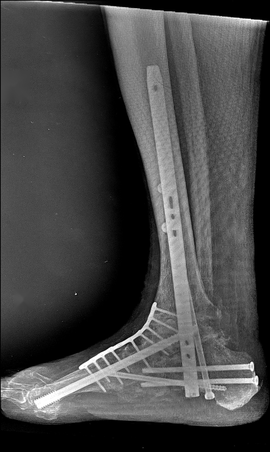
Weight-bearing X-rays of one of my reconstructions, in two planes.
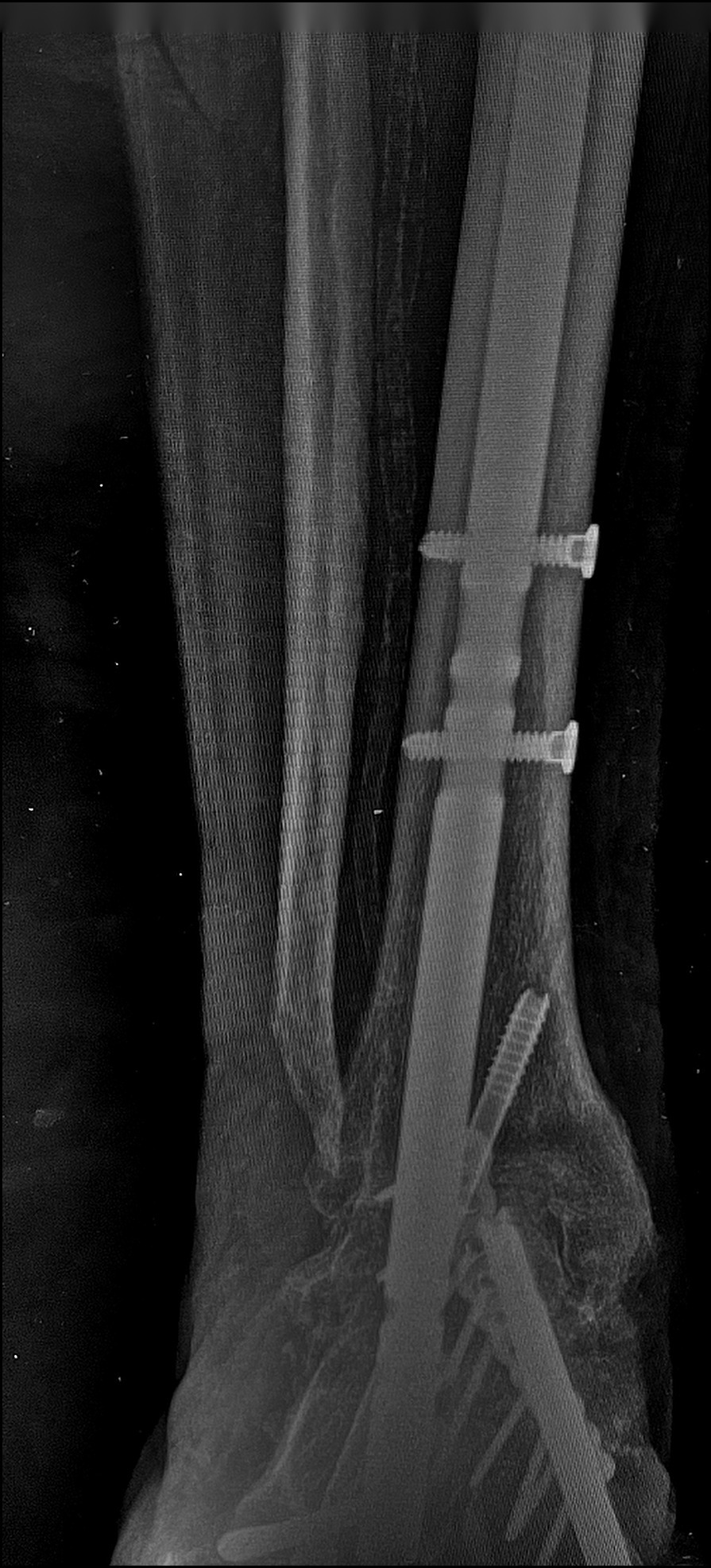
A nail passed up through the heel fuses the ankle and subtalar joints. In our published series of 70 reconstructions the hindfoot fused in 83%, and union was more likely when the nail filled the tibial canal and an additional compression screw was used (Bone & Joint Journal, 2022).
Long intramedullary beams and locking plates realign the inner and outer columns. An early single-bolt device was abandoned after an unacceptable failure rate in our first ten feet (Bone & Joint Journal, 2015).
About a third of limb-threatening deformities involve both hindfoot and midfoot. The hindfoot is corrected first, then the midfoot (Annals of Joint, 2023).
Two-stage reconstruction
When the foot is both deformed and infected, the infection is dealt with first and the reconstruction follows. This is the approach I presented at IADFS and will present in Padua in November 2026. The images below are from different patients.
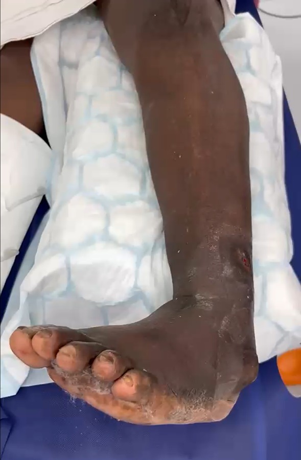
A collapsed midfoot, with the sole rolled over and skin breaking down over the bony prominence.
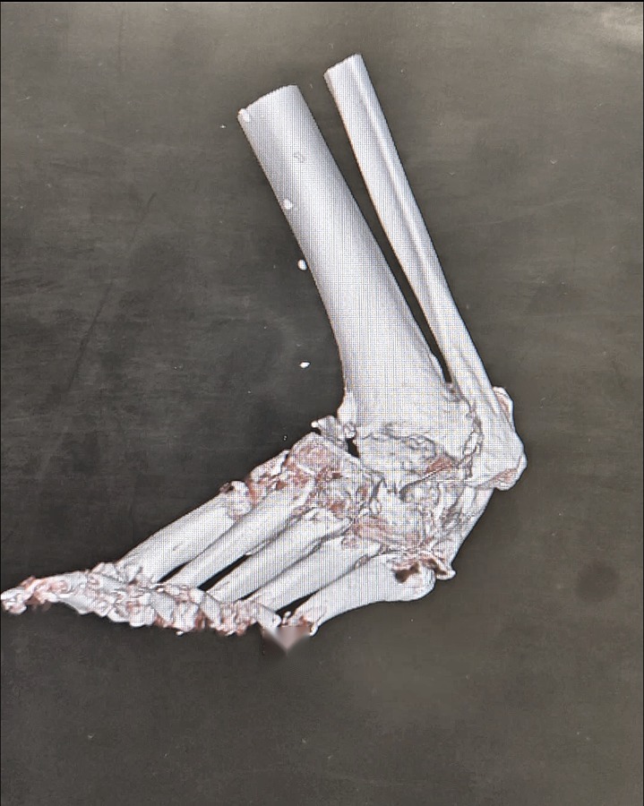
A 3D CT reconstruction shows the shape of the collapse and where the bone cuts need to be.
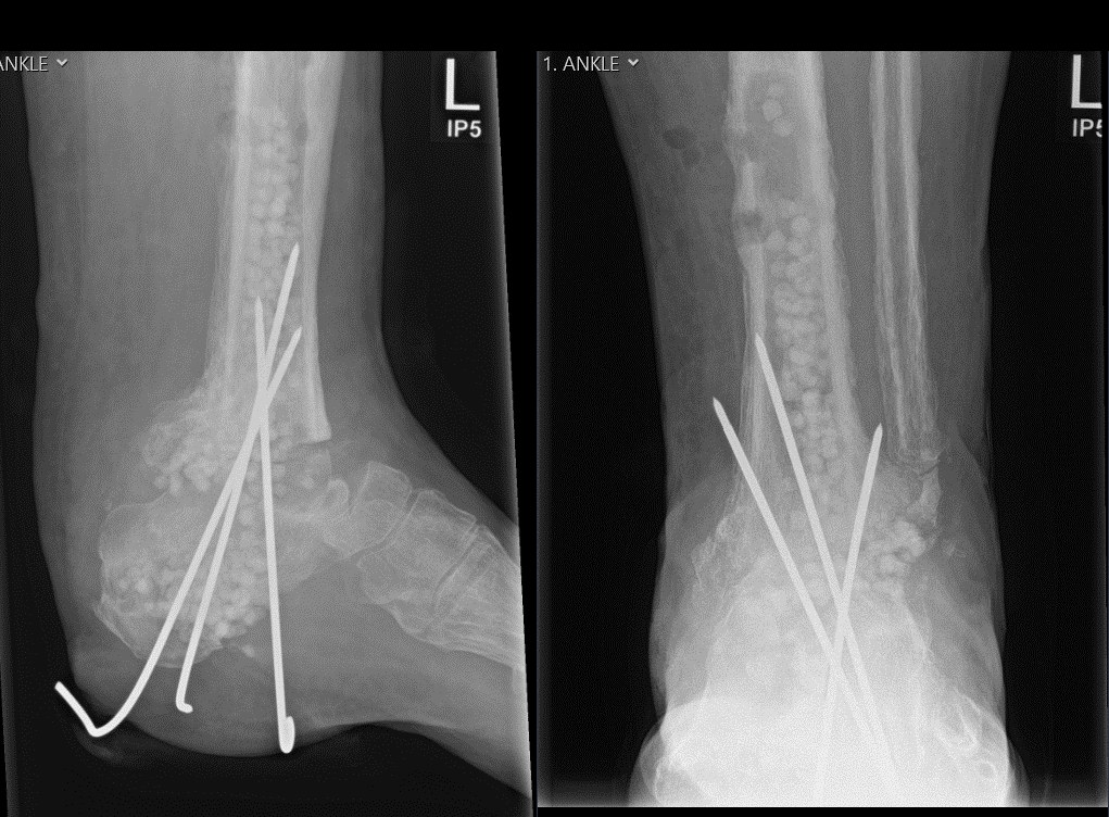
Debridement, deep samples, antibiotic-loaded bone graft substitute and temporary stabilisation with wires.
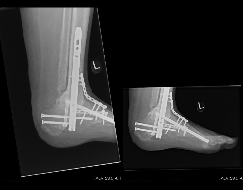
Definitive correction and long-segment fixation, once the inflammatory markers and the wound had settled.
Vascular assessment, ulcer and microbiology review, and imaging.
Antibiotics are adjusted to the cultures and continued until the blood markers and the wound are stable, normally six to ten weeks, with revascularisation or further debridement if needed.
Cast change at day three, then a total-contact cast for at least 12 weeks, with partial weight-bearing from 12 weeks once union is seen on X-ray or CT.
Published results from the unit: 23 infected, ulcerated feet treated in two stages, 83% fusion and 90% walking independently at a mean of 44 months (Kavarthapu & Budair, Bone & Joint Journal 2021).
Weight-bearing X-rays and CT, and 3D-printed models in complex cases.
Agreed with the diabetes team. Urgent cases need not always wait for an ideal HbA1c (Foot & Ankle Surgery, 2025).
A cast and strict offloading, then a boot, then surgical shoes or a brace.
Long-term podiatry and footwear review: the deformity is corrected, the neuropathy remains.